From February 16th - March 2nd, Dr. Philip Ferguson (Pathologist, Jonesboro, AR), Mike Tompkins (Leica, IHC and technical lab specialist, Phoenix, AZ), and Ronnie Vaca (Leica, histotechnologist and IHC specialist, Los Angeles, CA) worked on "Operation Kazuri Slide" in the histology laboratory to improve the H&E slide quality. Overall, the efforts were a huge success on all levels. The following blog post highlights the background and results of these efforts.
Background: The pathology laboratory has had limited quality H&E histology slides for many years. Most of the equipment in the laboratory was present at the time the lab opened at Kijabe in 1997, and they were well used at that time. Last summer a study was undertaken to identify the most important components of the slide quality with the intent to strategically target and improve the overall quality.
Project: From the results of the study from last summer, it was estimated that fixation (40%), microtomy (40%), and tissue processing (15%) were the three most important factors affecting slide quality. A plan was executed to improve each of these factors. Fixation has been a well established issue, in that the tissue has always "cut" differently than that in the United States. There has been a question with regards to the factors contributing to the fixation problem, and this was addressed by bringing in Powdered Formalin (BBC Biochemical) from the US along with installing a reverse osmosis water filtration system (sourced from Nairobi).
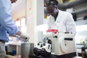
Microtomy quality consists of two main variables: (1) quality of the microtome and (2) experience/technique of the histotechnologist. Prior to this trip it was unknown if microtomy was primarily an issue of equipment or training. Mercedes Medical (Tampa, FL) donated a quality used Leica 2025 microtome through the efforts of Steve Westra (Statlab) and Brian Carson (Mercedes Medical). Inspection and preventive maintenance was performed prior to taking the microtome to Kijabe Hospital. Ronnie Vaca was also a key component to this trip given his vast experience as a histotechnologist to help install the microtome, train the laboratory staff, and assess their competence.
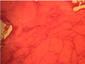
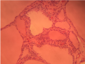
Results: After implementation of the Powdered Formalin and "new" microtome, the slide section quality quickly rose to the equivalent of a quality slide in the United States. Additionally, Phyllis and Peris proved themselves to be excellent histotechnologists who would be valued staff in any US histology laboratory. Ronnie also spent time educating on proper daily and routine maintenance on all of the histology equipment.
Conclusion:
- All future rotating pathologists will notice a significant improvement in histology slide quality. We also visited Lancet Labs and Aga Khan, and can confidently state the Kijabe slides are now of at least equal quality.
- The tissue when in a fixed state has the same texture and consistency as that in the US.
- It will be important that tissue is adequately fixed before going into the processor. A new protocol to allow cut sections in cassettes to fix an additional day for any tissue that is not well fixed at the time of grossing or thick sections. This was also felt to be an important process implementation during this onsite project.
- The laboratory staff were excited to see the slide quality improvements, and look forward to future projects.
Future Directions:
- H&E staining is still not optimized. This will be an ongoing project. Current protocol does not use a bluing solution. This has been ordered, and will be evaluated by future volunteers. The quality of the locally sourced stains are not known, and a comparison with stains from the US may be helpful if a future volunteer wants to take this project on.
- Immunohistochemistry - The laboratory staff was excited and highly motivated to bring in IHC stains for pathologist support and patient care. The assessment of all of the members of the onsite team was that the laboratory should be able to perform such testing at a high level of quality given the appropriate training and strategy.
- The onsite team plans to put together an IHC implementation plan in the next three months to present to the volunteer pathologists and Kijabe Hospital administration.
- H&E slide imaging -
- Leica has agreed to donate a slide imager to allow for remote consultations. This will be a great addition, especially given the improvements to slide quality.
- Unclear with regards to the time to implementation and shipping method
- Additional Microtome
- Leica has also agreed to donate a microtome. This will provide two quality functioning microtomes for the laboratory.
- Unsure of the time to implementation and shipping method.
- LIS System
- A need for a true pathology laboratory information system (LIS) for antomic pathology.
- Optimally, a cloud/internet based system would be optimal to supply reports to remote hospitals, allow for volunteer patholgists to finish reports from the US, and allow documentation for remote consultations. This could be particularly effective for cases where unstained slides may be brought back to the US for additional testing.